Human Anatomy and Physiology
Anatomical Planes of the Body
To understand anatomy of the human body, it has been divided and separated in few. The main planes and their sub-planes are following:
- Sagittal:Plane that runs down through the body, dividing the body into left and right portions. Subsections of the sagittal plane include:
- Mid-sagittalruns through the median plane and divides along the line of symmetry
- Parasagittal is parallel to midline but does not divide into equal left and right portions.
- Coronal (frontal):Plane that runs perpendicular to the sagittal plane and divides the body into anterior and posterior (front and back) portions.
- Transverse:Horizontal plane that divides the body into upper and lower portions; also called cross-section.
Directional Terms
In general, directional terms are grouped in pairs of opposites based on the standard anatomical position.
- Superior and Inferior. Superior means above, inferior means below. The elbow is superior (above) to the hand. The foot is inferior (below) to the knee.
- Anterior and Posterior. Anterior means toward the front (chest side) of the body, posterior means toward the back.
- Medial and Lateral. Medial means toward the midline of the body, lateral means away from the midline. Ipsilateralmeans on the same side—the left arm is ipsilateral (on the same side) to the left leg.
- Proximal and Distal. Proximal means closest to the point of origin or trunk of the body, distal means farthest away. Proximal and distal are often used when describing arms and legs. If you were describing the shin bone, the proximal end would be the end close to the knee and the distal end would be the end close to the foot. In the fingers of the hand, a proximal joint is closest to the wrist and a distal joint is farthest from the wrist.
- Superficial and Deep. Superficial means toward the body surface, deep means farthest from the body surface.
Other directional terms:
- Intermediate– means between—your heart is intermediate to your lungs.
- Caudal– at or near the tail or posterior end of the body.
- Visceral– may be used instead of deep.
There are also terms that describe specific body parts. Palmar describes the palm side of the hand. Dorsal describes the back side of the hand. Plantar describes the bottom of the foot.
Body Cavities:
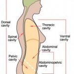
Body cavities are areas in the body that contain our internal organs. The dorsal and ventral cavities are the two main cavities. The dorsal cavity is on the posterior (back side) of the body and contains the cranial cavity and spinal cavity. In human anatomy, dorsal, caudal and posterior mean the same thing. The ventral cavity is on the front (anterior) of the body and is divided into the thoracic cavity (chest) and abdominopelvic cavity.
Dorsal Cavity
The dorsal cavity is further divided into subcavities:
- cranial cavity (also called the calvaria) which surrounds and holds the brain
- vertebral cavity (also called the spinal cavity) which includes the vertebrae (spinal column) and spinal cord
Ventral Cavity
The ventral cavity is on the front of the trunk. The diaphragm (the main muscle of breathing) divides the ventral cavity into two simple sub-cavities: thoracic and abdominal.
- Thoracic cavitywhich is surrounded by the ribs and chest muscles is superior (above) to the diaphragm and abdomen-pelvic cavity. It is further divided into the pleural cavities (left and right) which contain the lungs, bronchi, and the mediastinum which contains the heart, pericardial membranes, large vessels of the heart, trachea (windpipe), upper esophagus, thymus gland, lymph nodes, and other blood vessels and nerves.
- Abdomino-pelvic cavityis divided into the abdominal cavity and pelvic cavity. The abdominal cavity is between the diaphragm and the pelvis. It is lined with a membrane and contains the stomach, lower part of the esophagus, small and large intestines (except sigmoid and rectum), spleen, liver, gallbladder, pancreas, adrenal glands, kidneys and ureters. The pelvic cavity contains the bladder, some reproductive organs and the rectum.
The thoracic cavity is open at the top and the abdominal cavity is open at the bottom. Both cavities are bound on the back by the spine. Even though their location is defined, the shape of these cavities can change. How they change is very different. Breathing is the main way the shape of these two cavities changes. The abdominal cavity changes shape similar to a water-filled balloon. When you squeeze the balloon, the shape changes as the balloon bulges. When breathing compresses the abdominal cavity it “bulges” into a different shape. The abdominal cavity can also change shape based on volume—that is how much you eat and drink. The more you eat and drink, the harder it is for the diaphragm to compress the abdominal cavity—which is why it is harder to breathe after a large meal. Also, an increase in volume of the abdominal cavity decreases the volume in the thoracic cavity—you can take in less air. The thoracic cavity changes both shape and volume when you breathe. When you breathe out, the volume decreases; when you breathe in the volume increases. Because of how these two cavities are linked together in shape change, you can see that the quality of breathing affects the health of abdominal organs and the health of our organs affects the quality of our breathing.
Other Cavities
- Oral cavity – the space in the mouth inside the teeth and gums and is filled with the tongue when it is relaxed.
- Nasal cavity – in the nose
- Orbital cavities (left and right) – hold the eyes
- Middle ear cavities (left and right) – hold the small bones of the middle ear
- Synovial cavities – are inside the joint capsules that surround freely moving joints (such as the hip, knee, elbow, and shoulder)
Body Quadrants
Quadrants are another way our bodies are divided into regions for both diagnostic and descriptive purposes.
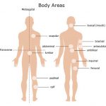
- Abdominal — relating to the abdomen. The abdomen is the part of the trunk between the chest and pelvis. It can be divided into three regions: the front, the belly; in back the loins; and on the sides, the flanks.
- Antecubital — region of the arm in front of the elbow
- Brachial — over the brachial artery in the upper arm
- Buccal — of or relating to the cheeks or the mouth
- Calf — of or relating to the calf
- Femoral — relating to the femur or thigh
- Inguinal — the groin or area in lower lateral regions of the abdomen
- Lumbar — area over the lumbar spine
- Popliteal — region on the back of the knee
- Scapular — of or relating to the area near the shoulder blade (scapula)
- Umbilical — relating to the central area of the abdomen near the bellybutton
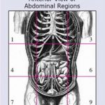
Body regions describe areas of the body that have a special function or are supplied by specific blood vessels or nerves. The most widely used terms are those that describe the 9 abdominal regions shown in the image to the right. The regions are named below and the corresponding regions are labeled 1-9.
- right (1) and left (3) hypochondriac regions– on either side of the epigastric region. Contains the diaphragm, some of the kidneys, right side of the liver, the spleen and part of the pancreas.
- epigastric region (2) – superior (above) the umbilical region and contains most of the pancreas, part of the stomach, liver, inferior vena cava, abdominal aorta and duodenum
- right (4) and left (6) lumbar (lateral) regions– on either side of the umbilical region. They contain portions of the large and small intestines and kidneys.
- umbilical region (5) – area around the umbilicus (belly button). Includes sections of the large and small intestines, inferior vena cava and abdominal aorta
- right (7) and left (9) iliac (inguinal) regions– are on either side of the hypogastric region and include portions of the large and small intestines.
- hypogastric (pubic) (8) region– inferior (below) the umbilical region. Contains parts of the sigmoid colon, the urinary bladder and ureters, the uterus and ovaries (women), and portions of the small intestines.
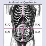
Quadrants are divide our bodies into regions for diagnostic and descriptive purposes. The quadrants are defined by drawing an imaginary line vertically (top to bottom) and horizontally (sideways) though the umbilicus (belly button). The following is a list of the organs in the four quadrants.
- Right Upper Quadrant (RUQ) – right lobe of liver, gallbladder, part of the transverse colon, part of pylorus, hepatic flexure, right kidney, and duodenum.
- Right Lower Quadrant (RLQ) – cecum, ascending colon, small intestine, appendix, bladder if distended, right ureter, right spermatic duct (men), right ovary and right tube and uterus if enlarged (women).
- Left Upper Quadrant (LUQ) – Left lobe of liver, stomach, small intestine, transverse colon, splenic flexure, pancreas, left kidney and spleen.
- Left Lower Quadrant (LLQ) – small intestine, left ureter, sigmoid flexure, descending colon, bladder if distended, left spermatic duct (men) left ovary and left tube and uterus if enlarged (women).


